
Endoscopic Management of Persistent Leak after Laparoscopic Sleeve Gastrectomy: A Case Report
by Melissa M. Beitner, MBBS; Jonathan Cohen, MD; and Marina S. Kurian, MD
Dr. Kurian and Dr. Beitner are from the Department of Surgery, NYU Langone Medical Center, New York, New York. Dr Cohen is from the Department of Medicine, Division of Gastroenterology, NYU Langone Medical Center, New York, New York.
FUNDING: No funding was provided.
DISCLOSURES: The authors report no conflicts of interest relevant to the content of this article.
Bariatric Times. 2012;9(2):22–24
ABSTRACT
Leaks after laparoscopic sleeve gastrectomy can be challenging to manage. Nonoperative management is preferred after the immediate postoperative period. No single treatment is effective in all cases. The best approach is to be persistent, to utilize a multidisciplinary team and to apply one or more endoscopic therapies, often in combination. We present a case of persistent leak after laparoscopic sleeve gastrectomy that highlights these issues.
INTRODUCTION
Staple line leaks after laparoscopic sleeve gastrectomy (SG) are associated with significant morbidity and even mortality. The reported incidence of leaks ranges from 0.5 to 5 percent.[1] In a systematic review of 24 studies with 1,749 patients, the incidence of leak was reported at an average of 2.7 percent.[2] Leak rates greater than 10 percent have been reported with revisional procedures.[3]
The clinical presentation of leaks ranges from asymptomatic radiographic findings to abdominal pain, peritonitis, and sepsis. Thus, a high index of suspicion is warranted.
Early leaks (those detected within a few days of surgery) may be amenable to primary surgical repair, but for late or persistent leaks, a nonoperative approach is preferred. A multidisciplinary team should be employed, utilizing one or a combination of open or percutaneous drainage of intra-abdominal collections, endoscopic clips, fibrin sealants, and self-expandable intraluminal stents.
Here, we present the case of a patient who developed a persistent leak and gastrocutaneous fistula after SG and was successfully managed with a combination of the aforementioned therapies.
Case report and management
A 36-year-old man with a history of obstructive sleep apnea (OSA) and morbid obesity (body mass index [BMI] 40.38kg/m2) underwent laparoscopic sleeve gastrectomy at an outside institution. The greater curve vessels were divided first and then the sleeve gastrectomy was started 5cm from the pylorus. The sleeve was created over a 40cm bougie. The staple line was buttressed with Seamguard (W.L. Gore and Associates, Inc, Flagstaff, Arizona) and the crossing staple lines were reinforced with 2-0 Vicryl sutures. An intraoperative methylene blue leak test was negative. A routine upper gastrointestinal (UGI) contrast series on Postoperative Day (POD) 1 showed a leak, and the patient returned to the operating room. Laparoscopic suture of a 1cm defect in the proximal staple line with debridement of the Seamguard, drainage of the intra-abdominal collection, and placement of a jejunal feeding tube were performed. Nutritional support, antibiotic therapy, and a proton pump inhibitor were instituted. A subsequent UGI series on POD 9 showed no further evidence of leak or obstruction. The patient then developed a left-sided pleural effusion that required drainage on POD 11 because of shortness of breath and a lowered oxygen saturation. On POD 13, the patient became febrile and tachycardic. Contrast-enhanced computed tomography (CT) of the abdomen and pelvis revealed ongoing leak from the gastric sleeve and a fluid collection in the left upper quadrant. Therefore, the patient again returned to the operating room for diagnostic laparoscopy, lysis of adhesions, aspiration of the abscess cavity, and placement of drains. A UGI series on POD 26 showed a small recurrent leak from the proximal suture line of the sleeve. Radiological drainage of an intra-abdominal collection was performed on POD 44 with a drainage catheter left in situ. Subsequent CT scans of the abdomen and pelvis on POD 50 and 63 to evaluate the abscess showed no discrete collection. However, the lateral left portal vein was thrombosed. The patient was placed on warfarin and discharged from hospital on POD 67.
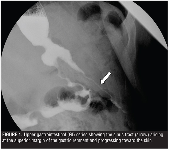
The leak continued to drain via a Jackson-Pratt (JP) drain with no pooling outside the drain on UGI contrast series. The patient subsequently developed a gastrocutaneous fistula with two tracts in communication with the gastric lumen (Figure 1), and during his outpatient follow up, the JP drain was removed. At this point, patient care was transferred to our practice. It had been five months since his initial surgery and his BMI had dropped from 40.38kg/m2 to 28.5kg/m2. We began management of the case by increasing his jejunal tube protein intake from 50g to 100g per day. The patient then underwent endoscopy with injection of 10mL of Tisseel (Baxter, Deerfield, Illinois) fibrin glue into the fistula tract. The tract had a 4mm wide opening and was located at an oblique angle in the dependent portion of a small pouch at the proximal aspect of the staple line below the gastroesophageal (GE) junction and above the sleeve (Figure 2
). The UGI contrast two weeks later showed no further leak and so the patient commenced a liquid diet.
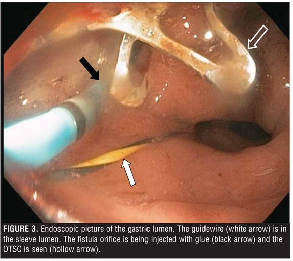
In the weeks following this, the patient presented with recurrent drainage from the gastrocutaneous fistula. This time the UGI contrast showed only one tract and so a second attempt at gluing was made. The tract remained patent. Endoscopic clip closure (of the leak) using an over-the-scope clip (OTSC) (Ovesco Endoscopy USA, Inc, Los Gatos, California) and dilation of the gastric sleeve to 18.5mm (using a 15-16.5-18mm x 180cm CRE balloon dilator [Boston Scientific, Natick, Massachusetts]) was attempted on two occasions as the patient declined stent placement (Figure 3
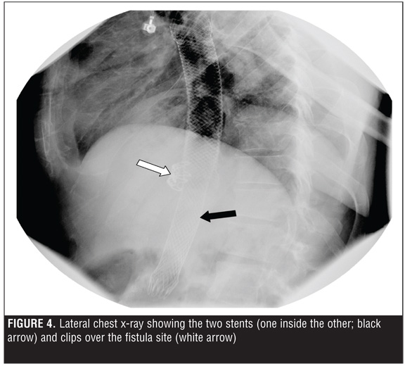
). During one attempt, the angulation of the tract orifice and its location in the dependent portion of a wider outpouching of mucosa impeded successful grasping of both sides of the tract and retraction into the cap on the tip of the scope prior to clip deployment. The tract persisted on UGI contrast series and was identified on endoscopy between the two clips in the area below the GE junction. The fistula tract was again injected with fibrin glue and a fully covered self-expanding metal stent (SEMS, Wallflex stent, Boston Scientific, Natick, Massachusetts]) was deployed. The mouth of fistula was mid-stent, and stent removal was planned in eight weeks. The patient experienced significant heartburn, nausea, and retching. On follow-up esophagram two weeks later, the length of the sinus tract was significantly reduced but the stent had migrated proximally. Therefore, a second SEMS was deployed. A partly covered Wallflex was utilized with the hope that the partly uncovered ends would help prevent migration. The proximal end of the second stent was located in the middle of the first stent and the distal end of the sleeve. As the uncovered proximal end was still within the lumen of the first fully covered stent, it was felt that removability of this second stent would not pose a problem. The area of the fistula, which was clearly demarcated by the metal clips, was tightly sealed in a position in the middle of the serial stent configuration (Figure 4
). The proximal end of the initial stent remained at 30cm from the incisors.
Following the second SEMS deployment, the fistula drainage stopped abruptly. The patient presented four weeks later with epigastric pain and vomiting. An esophagram showed proximal migration of the second SEMS; however, the tract had completely resolved (Figure 5
). The two stents were successfully removed together endoscopically as an outpatient after a total of eight weeks. His course post-stent removal was uneventful and he resumed an oral diet without clinical evidence of recurrent leak.
Discussion
Gastric leaks following SG present a complex management challenge. The widely held definition of an anastomotic leak is that proposed by the United Kingdom Surgical Infection Study Group: “the leak of luminal contents from a surgical join between two hollow viscera” or an outflow of gastrointestinal content through a suture line around an organ.[4] Leaks may be early (within 4 days of surgery), intermediate (between POD 5 and 9), or late (>10 days post operation).[4–6] They may also be classified as type I, those that are subclinical without evidence of dissemination, and type II, those that present clinically and show evidence of dissemination.[4,5] Leaks tend to occur at the GE junction.[7] According to Marquez et al,[4] leaks are more likely to occur in this area due to high intraluminal pressures and low compliance of the gastric sleeve, thus patients with distal stenosis are at greater risk. The pathogenesis of leaks is believed to be related to local ischemia near the staple line subsequent to the use of electrocautery in combination with higher intraluminal pressures postoperatively, rather than staple line dehiscence.[5,6] Baker et al[8] describe the etiology of leaks as being either mechanical/tissue causes or ischemic causes. Classic ischemic leaks occur 5 to 7 days postoperatively when wound healing is between inflammatory and fibrosis phases. These are less frequent. Thus, the majority of leaks are mechanical. This explains the variety of reinforcement techniques and materials available to attempt to reduce leak risk. Unfortunately, as a review by Chen et al[9] of 12 studies of 1589 SG cases shows, reinforcement of the staple line does not necessarily reduce leak rates.
Leaks can be asymptomatic but most commonly present with tachycardia.[10] In a series of 200 SG patients with a three-percent leak rate, Casella et al[11] report the most frequent symptoms to be abdominal pain, fever, vomiting, and dyspnea.
Diagnosis is made on imaging, with CT as the preferred imaging modality for diagnosis and UGI contrast series preferred for surveillance and for determining the origin of the leak;[12] however, CT may be unreliable in patients with a BMI greater than 50kg/m2.[13]
The basic tenets of management include nutritional support with correction of electrolyte imbalances, suppression of gastrointestinal secretions, bowel rest and elimination of downstream resistance, drainage of intra-abdominal collections, and infection and sepsis control. In the case of a gastrocutaneous fistula, skin protection around the external orifice of the fistula must also be considered. The management approach depends on timing of the leak; the tissue becomes inflamed as time from primary operation increases. This decreases the efficacy of both surgical repair and application of endoscopic clips.[14,15] There are several case series in the endoscopic literature using various clips, coagulation, tissue sealants, endoscopically placed sutures, and temporary covered stents either alone or in combination.[16–22]
This case illustrates the fact that multiple endoscopic modalities are often required, and that successful management may require repeated procedures. Prior to endoscopic intervention, the fistula tract should be mapped endoscopically and fluoroscopically, collections drained, and the intragastric mouth of the fistula debrided. Multiple endoscopic therapies might be considered in tandem or in combination. As attempts to seal or close the fistula may not work alone, as this case illustrates, strong consideration to diverting all secretions from the site using removable SEMS should be initially entertained.
Nonoperative management of a chronic fistula after laparoscopic SG requires a multimodal approach, persistence, and a range of endoscopic therapies. The bulk of the literature on endoscopic management of leaks after sleeve gastrectomy and other bariatric procedures comprises small case series in which a number of endoscopic therapies are used often in combination. There is currently no evidence-based algorithm for the treatment of leaks, including guidance as to which modality to try first. The literature does support the notion that treating physicians should be capable of offering the full range of available modalities. In the authors’ opinion, since multiple techniques are often required, strong consideration of using an initial combination of modalities appears reasonable for all but very small diameter fistulae.
References
1. de Aretxabala X, Leon J, Wiedmaier G, et al. Gastric leak after sleeve gastrectomy: analysis of its management. Obes Surg. 2011;21(8):1232–1237.
2. Brethauer SA, Hammel JP, Schauer PR. Systematic review of sleeve gastrectomy as staging and primary bariatric procedure. Surg Obes Relat Dis. 2009;5(4):469–475.
3. Ramalingam G, Anton CK. Our 1-year experience in laparoscopic sleeve gastrectomy. Obes Surg. 2011;21(12):1828–1833.
4. Marquez MF, Ayza MF, Lozano RB, et al. Gastric leak after laparoscopic sleeve gastrectomy. Obes Surg. 2010;20(9):1306–1311.
5. Burgos AM, Braghetto I, Csendes A, et al. Gastric leak after laparoscopic-sleeve gastrectomy for obesity. Obes Surg. 2009;19(12):1672–1677.
6. Csendes A, Braghetto I, Leon P, Burgos AM. Management of leaks after laparoscopic sleeve gastrectomy in patients with obesity. J Gastrointest Surg. 2010;14(9):1343–1348.
7. Lalor PF, Tucker ON, Szomstein S, Rosenthal RJ. Complications after laparoscopic sleeve gastrectomy. Surg Obes Relat Dis. 2008;4(1):33–38.
8. Baker RS, Foote J, Kemmeter P, et al. The science of stapling and leaks. Obes Surg. 2004;14(10):1290–1298.
9. Chen B, Kiriakopoulos A, Tsakayannis D, et al. Reinforcement does not necessarily reduce the rate of staple line leaks after sleeve gastrectomy. A review of the literature and clinical experiences. Obes Surg. 2009;19(2):166–172.
10. Hamilton EC, Sims TL, Hamilton TT, et al. Clinical predictors of leak after laparoscopic Roux-en-Y gastric bypass for morbid obesity. Surg Endosc. 2003;17(5):679–684.
11. Casella G, Soricelli E, Rizzello M, et al. Nonsurgical treatment of staple line leaks after laparoscopic sleeve gastrectomy. Obes Surg. 2009;19(7):821–826.
12. Tan JT, Kariyawasam S, Wijeratne T, Chandraratna HS. Diagnosis and management of gastric leaks after laparoscopic sleeve gastrectomy for morbid obesity. Obes Surg. 2010;20(4):403–409.
13. Jurowich C, Thalheimer A, Seyfried F, et al. Gastric leakage after sleeve gastrectomy-clinical presentation and therapeutic options. Langenbecks Arch Surg. 2011;396(7):981–987. Epub 2011 May 10.
14. Seebach L, Bauerfeind P, Gubler C. “Sparing the surgeon”: clinical experience with over-the-scope clips for gastrointestinal perforation. Endoscopy. 2010;42(12):1108–1111.
15. Albert JG, Friedrich-Rust M, Woeste G, et al. Benefit of a clipping device in use in intestinal bleeding and intestinal leakage. Gastrointest Endosc. 2011;74(2):389–397.
16. Papavramidis TS, Kotzampassi K, Kotidis E, et al. Endoscopic fibrin sealing of gastrocutaneous fistulas after sleeve gastrectomy and biliopancreatic diversion with duodenal switch. J Gastroenterol Hepatol. 2008;23(12):1802–1805.
17. von Renteln D, Denzer UW, Schachschal G, et al. Endoscopic closure of GI fistulae by using an over-the-scope clip (with videos). Gastrointest Endosc. 2010;72(6):1289–1296.
18. Eubanks S, Edwards CA, Fearing NM, et al. Use of endoscopic stents to treat anastomotic complications after bariatric surgery. J Am Coll Surg. 2008;206(5):935–938; discussion 938–939.
19. Merrifield BF, Lautz D, Thompson CC. Endoscopic repair of gastric leaks after Roux-en-Y gastric bypass: a less invasive approach. Gastrointest Endosc. 2006;63(4):710–714.
20. Nguyen NT, Nguyen XM, Dholakia C. The use of endoscopic stent in management of leaks after sleeve gastrectomy. Obes Surg. 2010;20(9):1289–1292.
21. Huang CS, Hess DT, Lichtenstein DR. Successful endoscopic management of postoperative GI fistula with fibrin glue injection: Report of two cases. Gastrointest Endosc. 2004;60(3):460–463.
22. Bege T, Emungania O, Vitton V, et al. An endoscopic strategy for management of anastomotic complications from bariatric surgery: a prospective study. Gastrointest Endosc. 2011;73(2):238–244.
Category: Case Report, Past Articles
Subscribe
If you enjoyed this article, subscribe to receive more just like it.


